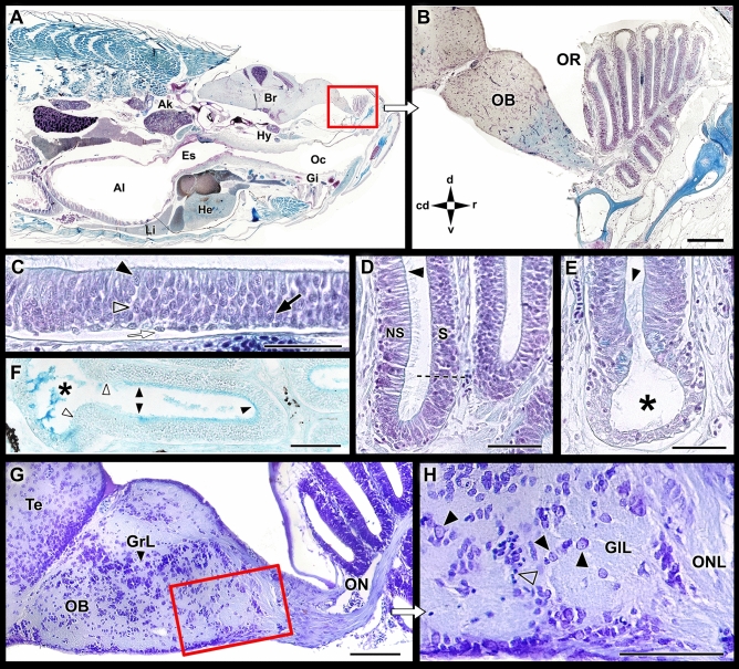
Figure 2.
Microscopic anatomy of the zebrafish olfactory system. (A) Low power sagittal section of the anterior zebrafish stained with Gallego’s trichrome. (B) Higher magnification of the inset in (A), showing the olfactory rosette and the olfactory bulb (OB). (C) Histological section of the olfactory sensory epithelium stained with Gallego’s trichrome. Black arrowhead, crypt cell; open arrowhead: microvillous cell; back arrow, ciliated cell; white arrow: basal cell. (D) Histological section of the medial side of the lamellae. The dotted line demarcates the nonsensory epithelium (NS) of the olfactory epithelium (S). Arrowhead, ciliated cells in the nonsensory epithelium. (E) The lateral rim of the lamellae-forming channel-like system (asterisk). Arrowhead, ciliated nonsensory cells. (F) Histological section of the lamellae stained by Alcian Blue. The luminal mucociliary complex is restricted to the sensory area (black arrowheads). The nonsensory epithelium border is free from acid mucins (white arrowheads), but Alcian Blue-stained secretions are concentrated inside the channel. (G) Sagittal section of the olfactory bulb. (H) Inset from (G) showing the olfactory nerve layer (ONL) and the glomerular layer (GlL). Black arrowhead, mitral cells; open arrowhead, periglomerular cells. Stains: (A–E) Gallego’s trichrome; (F) Alcian Blue; (G,H) Nissl stain. Ai, anterior intestine; Ak, anterior kidney; Br, Brain; Es, esophagus; Gi, gilts; GrL, granular layer; He, heart; Hy, hypophysis; Li, liver; Oc, oral cavity; OR, Olfactory rosette; Te, telencephalon; cd, caudal; d, dorsal; r, rostral; v, ventral. Scale bars: 100 µm (B,G,H); 50 µm (C–F).

