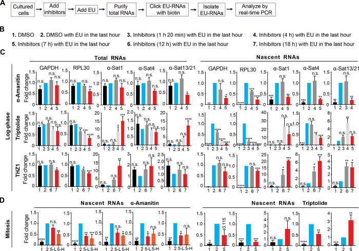
Figure 1.
Transcriptional inhibitors exhibit distinct, even opposing, efficacies on the suppression of ongoing α-satellite and gene transcription. (A) A flow chart illustrating the preparation and analysis of EU-labeled nascent RNAs. Cultured cells are referred to as log-phase (C) or nocodazole-arrested mitotic (D) cells. (B) Experimental conditions that were used in C. The times listed here refer to the durations of inhibitor treatment. The inhibitor concentrations were selected based on the toxicity of each inhibitor on the tested human cells. They were near but lower than lethal concentrations. No more than 20% of dead cells were observed under each of the following conditions: α-amanitin: 50 µg/ml, triptolide: 1.4 µM, flavopiridol: 1.0 µM, and THZ1: 120 nM. Treatment of these inhibitors seemed not to significantly alter the cell cycle profile of cultured log-phase (C) or mitotic (D) cells. EU was usually added 1 h before cell harvest. (C) HeLa Tet-On cells were treated with various types of transcriptional inhibitors as described in B. (D) Nocodazole-arrested mitotic HeLa Tet-On cells were treated with DMSO, α-amanitin (5-L, 5 µg/ml; 5-H, 50 µg/ml) or triptolide for 7 h with EU treatment in the last hour. EU-RNAs were prepared and analyzed by real-time PCR. The details are recorded in the Materials and methods. The average and standard error calculated from at least three independent experiments are shown here. *, P < 0.05; **, P < 0.01; ***, P < 0.001; ****, P < 0.0001. α-Sat, α-satellite.

