
Figure 6.
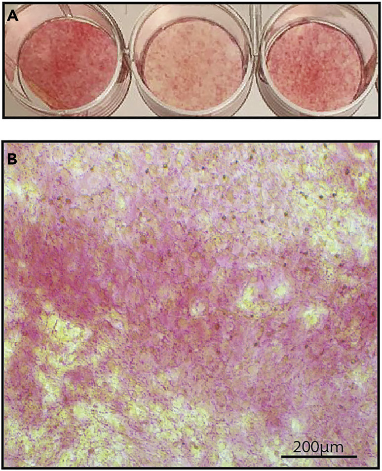
Sirius red staining
Representative image of osteoblasts after Sirius red 17 days after seeding the cells visualized using a standard camera (A) or under light microscope (B; 10×, Scale: 200 μm).
Official websites use .gov
A
.gov website belongs to an official
government organization in the United States.
Secure .gov websites use HTTPS
A lock (
) or https:// means you've safely
connected to the .gov website. Share sensitive
information only on official, secure websites.
Sirius red staining
Representative image of osteoblasts after Sirius red 17 days after seeding the cells visualized using a standard camera (A) or under light microscope (B; 10×, Scale: 200 μm).