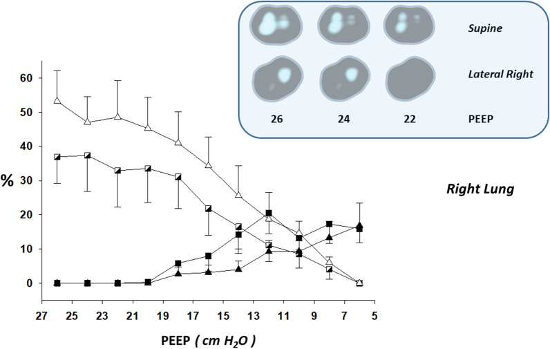
Fig. 3.
Lung collapse and overdistension by electrical impedance tomography in supine vs. targeted lateral body position within the right lung. Left-to-right lung asymmetry was present on initial X-Ray taken in supine body position: unequivocally more opacities within the left lung. Thus lateral right positioning (30°) was indicated (“targeted”) and performed with the platform-based rotation bed Multicare® (LINET). Line graphs of electrical impedance tomography (EIT)-based estimations of collapse and overdistension during decremental positive end-expiratory pressure (PEEP) titrations (supine vs. targeted lateral body position) are shown (mean ± SEM). Some illustrative and representative EIT images of overdistension are also shown: overdistended pixels in white. Note the asymmetric distribution of overdistension between the right and left lungs (concentration and predominance of overdistension within the right lung); and that the amount of overdistended units within the right lung in the supine body position was minimized in the lateral right one. Also note that the regional distribution of overdistension in the supine body position was much less gravitational-dependent than it is usually present in “typical” acute respiratory distress syndrome. X axis: Decremental PEEP levels of the EIT-PEEP titrations. Y axis: Percent of overdistended and collapsed lung units out of the total lung imaged by EIT. Triangle: Supine body position. Square: Targeted lateral body position (lateral right). Black triangle and black square: Percent of collapsed lung units out of the total lung imaged by EIT. White triangle and white semi-filled square: Percent of overdistended lung units out of the total lung imaged by EIT

