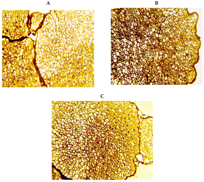
Figure 6.
Silver staining for transverse sciatic nerve specimens. Images show well organized nerve fiber axons, including well-structured myelin sheathes enclosed by endoneurium in the saline group (A), profound staining by silver stain and a substantial decrease in the myelin sheathes in the diabetic group (B), less degeneration in the myelin sheathes and reduced silver staining in the DM+memantine group (C). Silver stain ×400.

