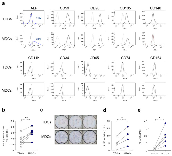
Figure 1.
ALP was highly expressed in MDCs compared with TDCs. (a) Tendon and muscle surface markers such as ALP, CD59, CD90, CD105, CD146, CD11b, CD34, CD45, CD74, and CD164 were analyzed by FACS. (b) Quantification of surface ALP expression (n = 6). (c) All TDCs and MDCs were cultured in growth medium for a day and cells were assessed with ALP staining (scale bar = 200 µm, (d) ALP activity (n = 4), or (e) qPCR for ALP mRNA expression (n = 5). *, p < 0.05; **, p < 0.01. Data are presented as the mean ± SEM.

