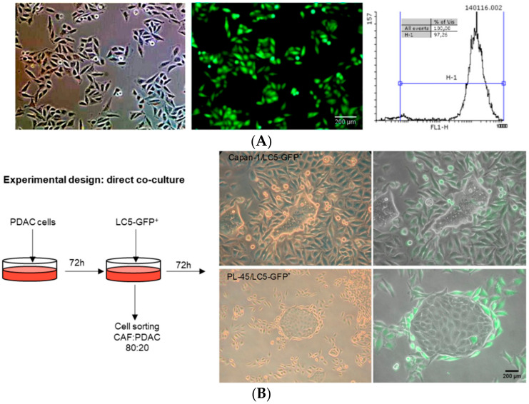
Figure 1.
Photographs of phase contrast and fluorescence microscopy of the selection of fibroblast LC5-GFP+ after FACS sorting. The percentage of GFP expressing fibroblasts was higher than 97% (A). Strategy scheme of direct cocultures: representative images at 72 h of pancreatic tumor cells (Capan-1 or PL-45) and fibroblast LC5-GFP+ (B). Green cells are GFP-transfected fibroblasts.

