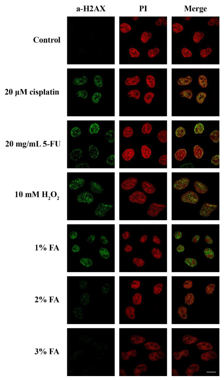
Figure 3.
Stress agents and inefficient fixation induced DNA damage. Fluorescent patterns of H2AX in HeLa cells treated with 20 µM cisplatin for 24 h, 20 µg/mL 5-FU for 48 h, 10 mM H2O2 for 1 h, or fixed for 5 min with 1%, 2%, or 3% FA. H2AX was detected using the anti-phospho-histone H2AX (Ser139) monoclonal antibody (Millipore), while nuclei were stained with PI. Scale bar: 10 µM.

