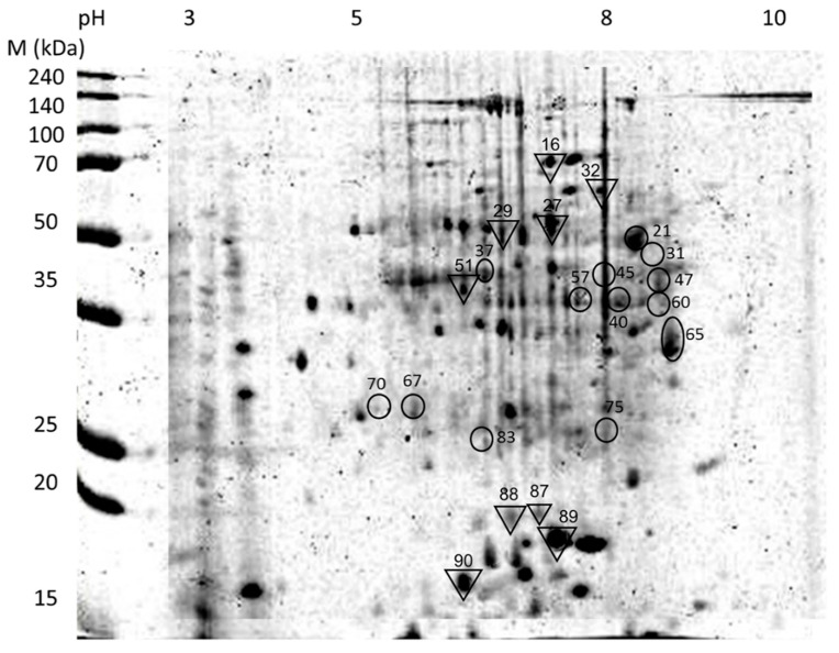
Figure 5.
Two-dimensional electrophoresis merged master gel of separated total proteins from the fungus–termite interaction. Protein spots with significant expression are identified in Coptotermes curvignathus (circles) and Metarhizium anisopliae (reversed triangles). Numbers shown next to the protein spots are their respective protein IDs.

