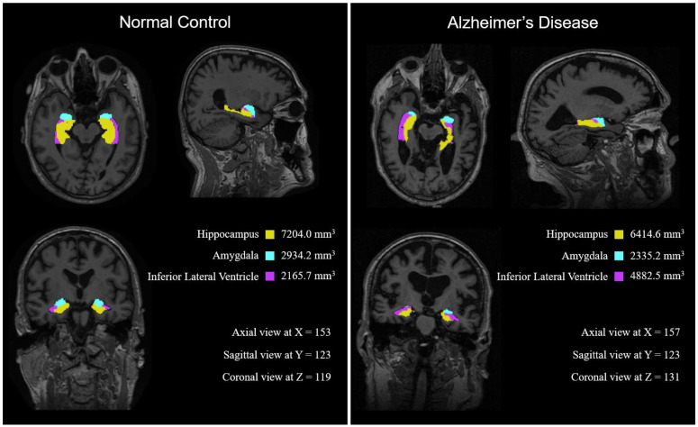
Figure 6.
Two representative MRI images from the NC and AD groups. The three areas identified are marked as follows: Hippocampus: Yellow, Amygdala: Cyan, and Inferior Lateral Ventricle: Purple). Notably, the NC’s hippocampus and amygdala are larger and the inferior lateral ventricle is smaller than that of the AD subject.

