Figure 2.
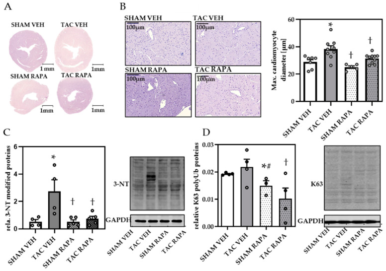
Rapamycin prevented TAC-induced cardiac remodeling in failing hearts. (A) H&E staining of cardiac cross-sections (magnification 1×, scale bar = 1 mm). (B) Representative images of H&E stained cardiomyocytes (magnification 20×, scale bar = 100 µm) and quantification of max. cardiomyocyte diameter. (C) Relative 3-nitrotyrosine (3-NT) quantification and representative immunoblot. (D) Relative K63-polyubiquitinated protein (K63) quantification and representative immunoblot. Immunoblot data are normalized to GAPDH and all data are presented as Mean + SEM of biological replicates. Statistical analyses were performed by two-way-ANOVA followed Tukey’s posttest or unpaired t- test (indicated with #). Statistically significant differences are shown by p < 0.05, * vs. SHAM VEH, † vs. TAC VEH.

