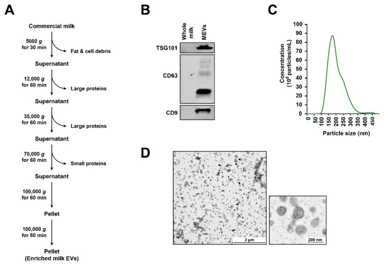
Figure 1.
Characterization of milk-derived extracellular vesicles (MEVs). (A) Schematic diagram of MEVs isolation. (B) Western blotting analysis of 30 µg of MEVs and whole milk probed with TSG101. (C) Nanoparticle tracking analysis of MEVs with the highest peak at 170 nm. (D) Transmission electron miscopy (TEM) images suggested the presence of vesicles in the preparation.

