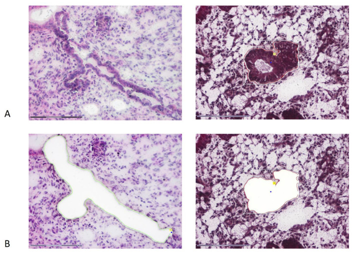
Figure 1.
A single representative endometrial gland before (A) and after (B) laser microdissection from 8 µm tissue slice. For a demonstration of gland mapping, eosin and hematoxylin staining was used on this sample (on the left). For DNA extraction, sections were stained with hematoxylin only (on the right).

