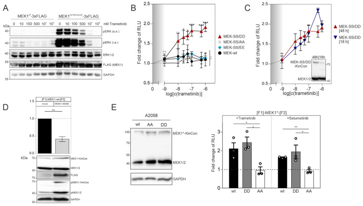
Figure 2.
MEKi and BRAF activities affect MEK1 KinCon reporter dynamics: (A) ERK1/2 activation following overexpression of indicated MEK1 fusion proteins and exposure to increasing doses of the MEKi trametinib for 1 h. Shown is a representative Western blot of n = 4 independent experiments. Antibodies directed against phosphorylated ERK (pERK, either short exposure (s.e.) or long exposure (l.e.)), total ERK1/2, flag or GAPDH as loading control were used (HEK293T). (B) Dose-dependent effect of 1 h trametinib treatment on the dynamics of indicated MEK1 KinCon reporters. Data points represent obtained bioluminescence signals in RLU relative to the DMSO control. They were plotted against increasing inhibitor concentrations on a logarithmic scale (± SEM from n = 8 independent experiments, HEK293T). (C) Shown are the dose-dependent responses of trametinib exposure upon expression of the MEK1 KinCons for 18 h (n = 3) versus 48 h (n = 8) (HEK293T). (D) Bars represent obtained bioluminescence signals in RLU relative to the signals of the MEK1 KinCon reporter in the presence of overexpressed and flag-tagged BRAF-V600E (±SEM from n = 5 independent experiments; normalized on KinCon reporter expression levels). Antibodies directed against phosphorylated MEK1/2, total MEK1/2, flag and GAPDH were used (HEK293T). (E) A2058 transiently expressing indicated KinCons were subjected to MEKi exposure with 1 µM trametinib, selumetinib or DMSO for 1 h. Bioluminescence signals in relative light units (RLU) relative to the signals of the DMSO treatment are depicted (± SEM from n = 3 independent experiments). Student’s t-test was used to evaluate statistical significance. Confidence level is indicated by asterisk as: * p < 0.05, ** p < 0.01, *** p < 0.001.

