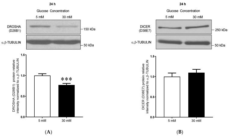
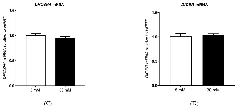
Figure 1.
High glucose treatment decreases DROSHA protein expression in human primary dermal microvascular endothelial cells (HDMECs). (A,B) Representative immunoblots and densitometry analysis for (A) DROSHA (D28B1) and (B) DICER (D38E7) protein expression levels in primary human dermal microvascular endothelial cells (HDMEC) after 24 h treatment with low (5 mM) or high (30 mM) glucose concentrations. α/β-TUBULIN was used as a loading control. Data are means ± SEM. (n = 12, t-test, *** p < 0.001 vs. 5 mM). (C,D) DROSHA and DICER1 mRNA expression relatively to HPRT in HDMEC treated with low (5 mM) or high (30 mM) glucose concentrations for 24 h. Data are means ± SEM. (n = 8, DICER1 mRNA; n = 12, DROSHA mRNA).


