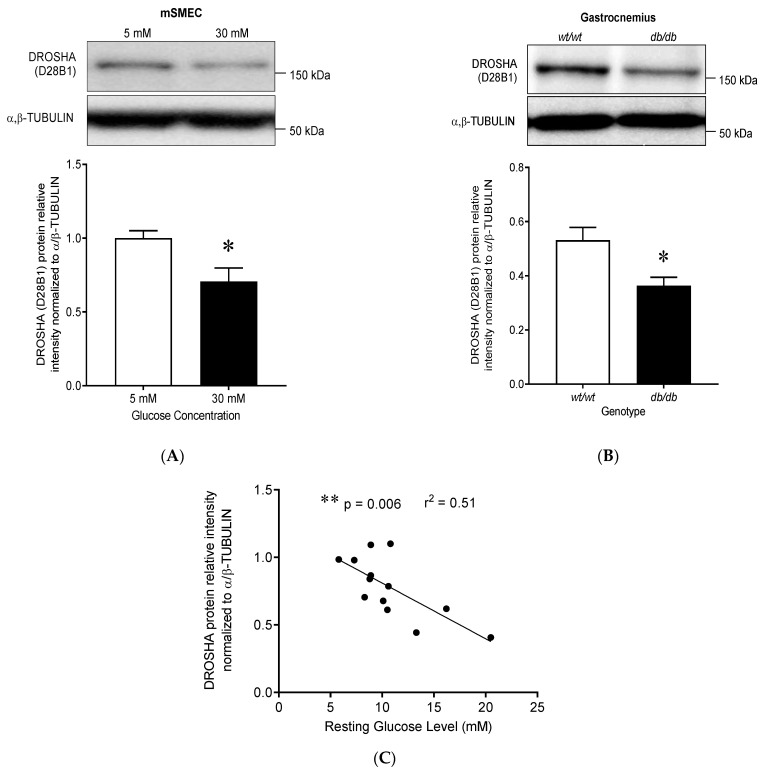
Figure 5.
High glucose conditions decrease DROSHA protein expression in mouse skeletal muscle tissue. (A) Immunoblot of DROSHA expression in mouse primary endothelial cells isolated from skeletal muscle tissue (mSMECs) following 24 H treatment with high (30 mM) or low (5 mM) glucose concentrations. α/β-TUBULIN was used as a loading control. High glucose concentration (30 mM) decreased DROSHA protein expression (data are means ± SEM, n = 6, * p < 0.05). (B) Immunoblot of DROSHA expression in gastrocnemius muscles from wild-type (wt/wt) and leptin receptor deficient (db/db) mice (data are means ± SEM, n = 4–7, p = 0.034). The db/db mice expressed significantly less DROSHA protein compared to their wild-type littermates (data are means ± SEM, n = 4–7, * p < 0.05). (C) Correlation analysis between DROSHA protein expression and resting blood glucose levels in gastrocnemius muscles from 4-week-old mice, including 2 wt/wt, 2 db/db and 9 db/wt mice. Resting blood glucose is negatively correlated with DROSHA protein expression in mice skeletal muscle (n = 13, ** p = 0.006).

