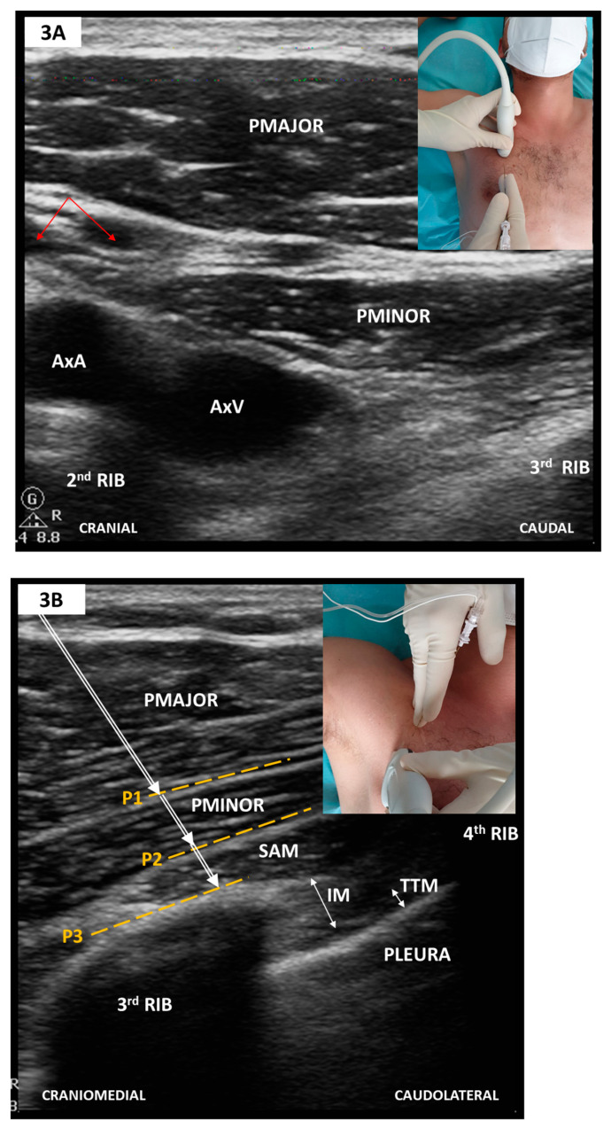
Figure 3.
(A) Parasagittal scan along the medioclavicular line-2nd rib level; (B) Oblique scan after a slight medial tilt with inferolateral sliding towards the midaxillary line-4th rib level (see text). PMAJOR, pectoralis major muscle; PMINOR, pectoralis minor muscle; AxA, axillary artery; AxV, axillary vein; red arrows, thoracoacromial artery and vein; SAM, serratus anterior muscle; IM, intercostal muscle; TTM, transversus thoracic muscle; P1, PECS I plane; P2, superficial plane for SAPB/PECS II; P3, deep plane for SAPB/PECS II. To elicit an adequate SAPB coverage, P2 or P3 need to be targeted at the 4th or 5th rib level.

