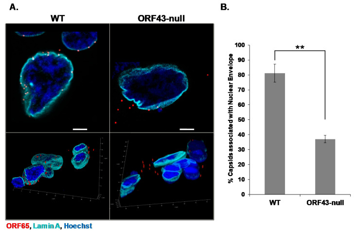
Figure 2.
Open reading frame (ORF) 43-null Kaposi’s sarcoma-associated herpesvirus (KSHV) capsids display relatively lower extent of association with the nuclear envelope compared to wild-type (WT) capsids. SLK cells were infected with WT or ORF43-null KSHV BAC16 virions. Six hours post-infection, cells were fixed, and capsids were detected with an antibody to the small capsid protein ORF65 and a Rhodamine-conjugated anti-mouse secondary antibody (red), while the nuclear envelope was observed using an antibody to Lamin A and an Alexa Fluor 647-conjugated anti-rabbit secondary antibody (cyan). Three-dimensional images were acquired by Z-stack with the super Z galvanometer substage, and 3D reconstruction with LASX software (Leica) (A). 1330 cells, containing 7890 WT capsids, and 2231 cells, containing 4290 ORF43-null capsids were counted in three independent experiments. The percentage of WT and ORF43-null capsids associated with the nuclear envelope is presented. Data are expressed as mean ± SD. ** p = 0.0023 (p < 0.01) (B). Scale bars = 5 µm.

