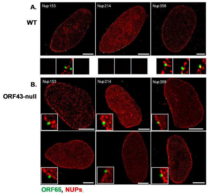
Figure 4.
Co-localization of KSHV WT and ORF43-null capsids with Nup358, Nup214, and Nup153 detected by stimulated emission depletion (STED) microscopy. U2OS cells were infected with WT or ORF43-null KSHV. Six hours post-infection, cells were treated with 0.1% Triton X-100 for 5 min, to remove the cytoplasm, and fixed in 4% para-formaldehyde in PBS for 20 min. Capsids were detected with an antibody to ORF65 and a secondary anti-mouse conjugated to Alexa 488 (green), and co-localization with Nup214, Nup153, or Nup358 was examined with Nup-specific antibodies, and secondary anti-rabbit Cy3-conjugated antibodies (red); WT capsids (A), ORF43-null capsids (B). Scale bars = 5 µm.

