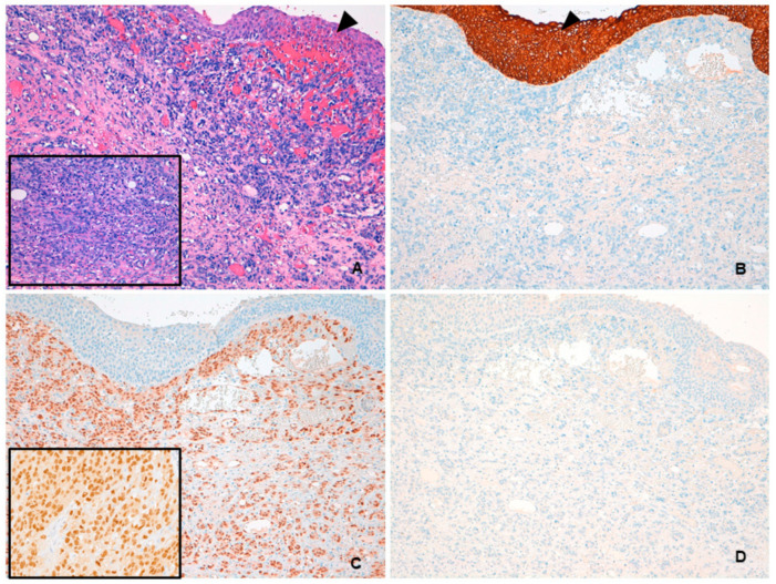
Figure 1.
(A) Hematoxylin and eosin low-power image showing angiosarcoma with diffuse infiltration of the lamina propria. Overlying urothelium is uninvolved (arrowhead). At higher magnification, short fascicles of atypical spindle cells with slit-like vascular spaces were evident (inset). (B) PAN cytokeratin AE1-AE3 immunoreactivity was seen in the urothelium (arrowhead) but not in neoplastic cells. (C) Angiosarcoma stained positively for ERG, with nuclear localization (inset). (D) HHV-8 was not expressed by tumor cells.

