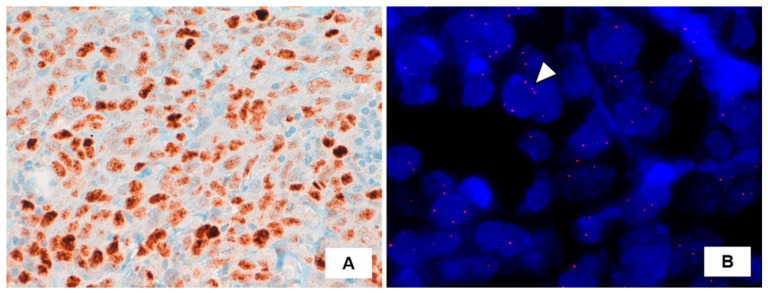
Figure 2.
(A) Immunohistochemistry for c-MYC protein displaying intense expression in angiosarcoma cells (brown precipitate indicates the presence of the target antigen; hematoxylin counterstaining). (B) Fluorescence in situ hybridization (FISH) results using a SpectrumOrange LSI c-MYC probe (Vysis, Abbott Park, IL, USA). Representative tumor cell (arrowhead) showing no increase in the number of signals, indicative for absence of MYC locus amplification. Nuclei are counterstained with 4–6-diamino-2phenylindole (DAPI, blue).

