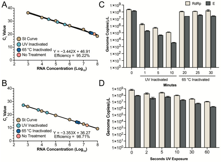
Figure 2.
Detection of RNA genomes following inactivation. Inactivated viral supernatants were subjected to RNA extraction and detection. (A,B) A standard curve was produced for the RdRp (A) and E (B) assays using serial diluted in vitro transcribed RNA (brown circles). RNA from UV and heat inactivated samples (light blue and dark blue, respectively) fall within the linear detection of both assays. The untreated genomic RNA is represented by the pink circle. (C) Quantification of the untreated RNA, UV inactivated samples at 1, 5 and 10 min and heat inactivated samples at 20, 25 and 30 min are shown. The mean and SEM from triplicate wells for the RdRp (tan) and E (grey) assays are shown. Samples inactivated via UV-C exposure for 1, 5, or 10 min were exposed to 4 × 10⁵, 2 × 10⁶, and 4 × 10⁶ µJ/cm2, respectively. (D) Quantification of untreated RNA and UV inactivated samples at 2, 5, 10, 30 and 60 s for the RdRp (tan) and E (grey) assays.

