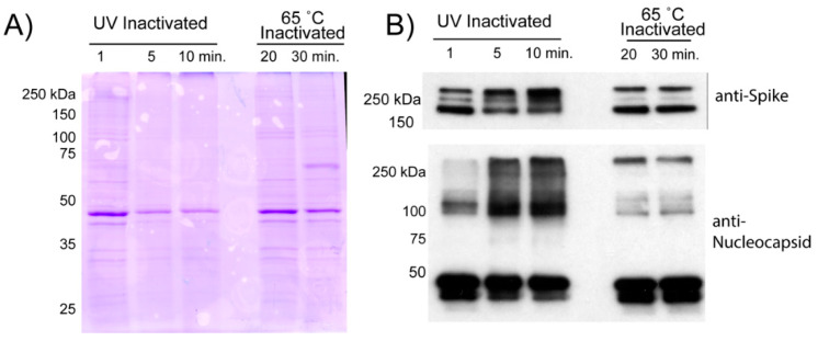
Figure 3.
Comparison of virion protein quality following inactivation. Viral supernatants inactivated by UV or Heat exposure were loaded onto a two-step sucrose gradient and subjected to ultracentrifugation. The resulting pellets were resuspended in PBS and analyzed for protein content. (A) 5 µg of resuspended pellets from the two inactivation conditions were loaded onto a 10% SDS-PAGE gel that was then subjected to Coomassie staining. (B) 0.2 µg of each pellet was run on a 10% gel and transferred to PVDF membrane for Western analysis for SARS-CoV-2 Spike or Nucleocapsid. Protein extracted from SARS-CoV-2 infected cells was run as a positive control.

