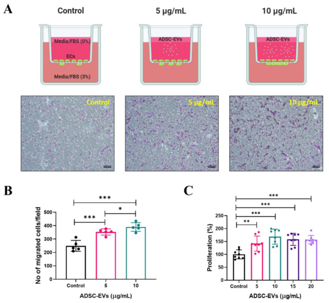
Figure 4.
Increased migration and proliferation of endothelial cells by treatment with ADSC-EVs. (A) Graphical scheme of the migration assay (figure created with BioRender.com) and phase contrast microscopy images of migrated endothelial cells after 24 h treatment with ADSC-EVs (5 and 10 µg/mL). (B) Quantified data of migrated cells shown in (A) (n = 5). (C) Graph showing the proliferation of endothelial cells quantified via CCK8 assay after 24 h treatment with 0–20 µg/mL of ADSC-EVs (n = 8). * p < 0.05, ** p < 0.01, *** p < 0.001 by Student’s t-test.

