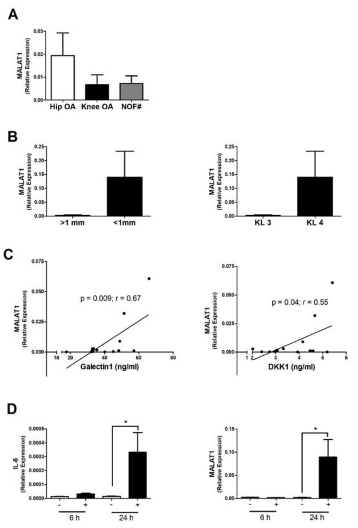
Figure 1.
Expression of MALAT1 in subchondral bone and OA osteoblasts. (A) Relative expression of MALAT1 in the subchondral bone tissue of end-stage hip OA patients (n = 9) compared to end-stage knee OA patients (n = 8) and non-OA neck of femur fracture (NOF#) patients (n = 6). MALAT1 expression was determined by qPCR and normalised to 18S. Bars represent mean expression ± SEM. (B) Relative expression of MALAT1 in the subchondral bone tissue between OA patients with more severe radiographic signs of OA (n = 13) with joint space < 1 mm and KL grade 4, compared to those with less severe joint damage (n = 4) with joint space > 1 mm and KL grade 3. (C) Correlation between subchondral bone expression of MALAT1 and the serum concentration of Galectin 1 (ng/mL) and DKK1 (ng/mL) in OA patients (n = 17). (D) Effect of IL-1β stimulation (1 ng/mL) upon the expression of IL-6 and MALAT1 in primary OA osteoblasts at 6 h and 24 h. * = p < 0.05, compared to non-stimulated control.

