Figure 4.
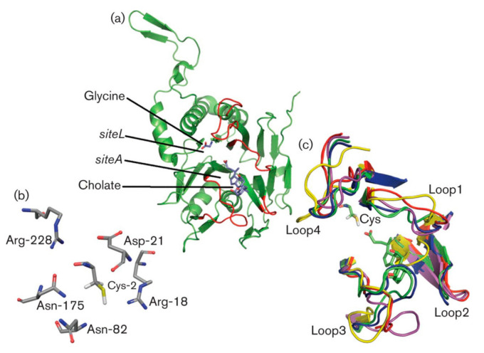
(a) The 3D structure of Clostridium perfringens CpBSH following the hydrolysis of glycocholic acid (GCA) with the products glycine bound in the active site (siteL) and cholate bound in the binding site (siteA) (both products are shown in stick representation and labelled). (b) The geometrical arrangement of the six major catalytic residues in the active site of CpBSH. (c) The superimposition of the four loops of the substrate binding site (loop1-loop4) from Bifidobacterium longum BlBSH (red), CpBSH (magenta), Bacteroides thetaiotaomicron BtBSH (yellow), Lyinibacillus sphaericus BspPVA (blue) and Bacillus subtilis BsuPVA (green). The active site nucleophilic residue Cys is shown and labelled. From [10].

