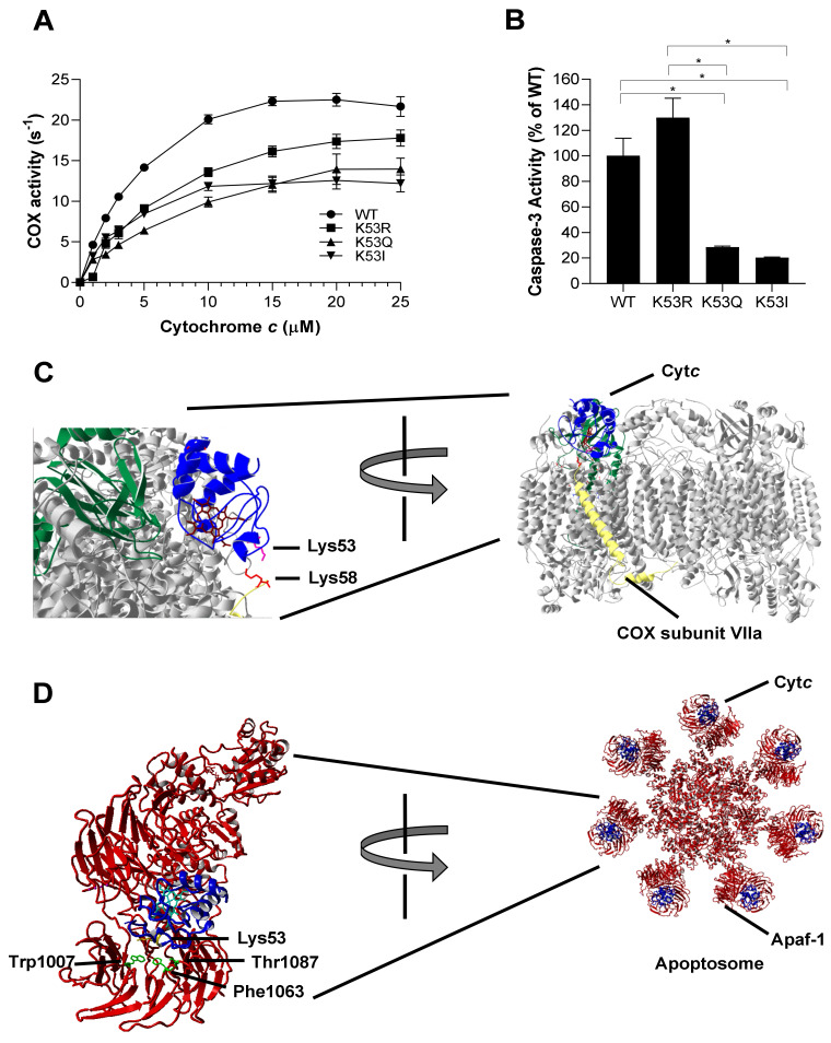
Figure 2.
(A) Oxygen consumption rate was measured in the presence of 30 nM of bovine liver COX using an Oxygraph system. WT, K53R, K53Q, and K53I Cytc variants were titrated at concentrations of 0, 1, 2, 3, 5, 10, 15, 20, and 25 µM (n = 3). Data are represented as means±SEM. (B) Cytosolic extracts of HeLa cells were incubated with recombinant WT, K53R, K53Q, and K53I variants for 2.5 h at 37 °C. Fluorescence that resulted from caspase-3 mediated cleavage of the rhodamine substrate DEVD-R110 was used as a measure of caspase-3 activity (n = 3). Data are represented as means ± SEM, * p < 0.05. (C) Docking model of COX and Cytc [32] showing the interaction of Lys53 residue of Cytc with Lys58 residue of COX subunit VIIa within 5 Å. (D) Cytc Lys53 interaction with Apaf-1 represented on the human apoptosome structure [33]. Apaf-1 residues Phe1063 (within 5 Å) and Trp1007 and Thr1087 (within 6 Å) are in close proximity to Lys53 residue.

