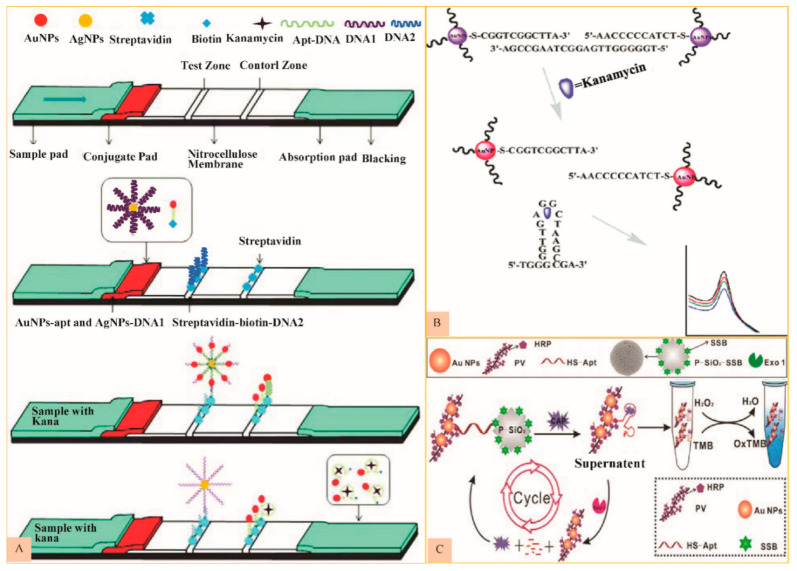
Figure 3.
(A) Schematic figure showing system alignment and detection principle of strip aptasensor for KAN (Reproduced with permission from [65] © 2018 Royal Society of Chemistry). (B) Schematic diagram to represent spectrophotometric kanamycin detection (Reproduced with permission from [66] © 2014 Royal Society of Chemistry). (C) Scheme depicting the suggested biosensing of STR dependent on the developed colorimetric aptasensor (Reproduced with permission from [72] © 2017 Elsevier).

