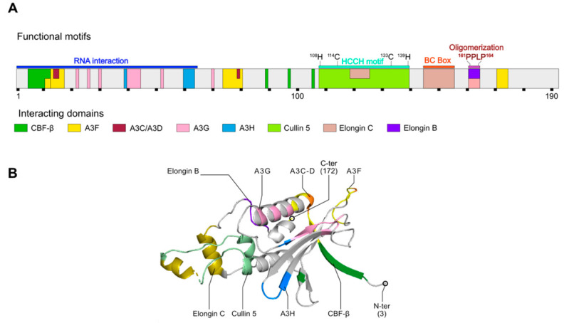
Figure 2.
HIV-1 Vif protein structure and interactions with E3-ubiquitin ligase complex members. (A) Vif mono-dimensional structure (Genbank M19921; UniProtKB—P12504 (Vif_HV1N5)) is represented using 10 amino acids per scaling unit (black squares under the structure). Functional motifs are represented with colored rectangles above the structure. Interaction regions with A3 proteins or members of the E3-ubiquitin ligase are represented in color inside the structure. (B) Vif 3D structure (PDB: 4N9F; positions 3-172) is represented in ribbons. N- and C- terminal extremities (N-ter and C-ter, respectively) as well as interaction regions with several partners are colored and annotated on the structure. Adapted from [81].

