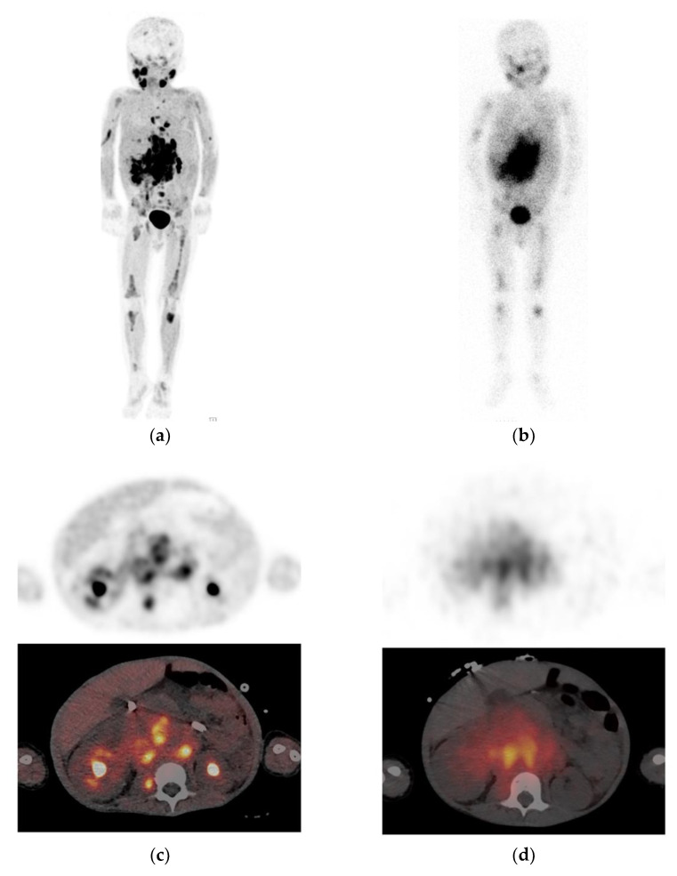
Figure 2.
Example of a neuroblastoma patient with extensive metastasis who underwent [123I]mIBG and [18F]mFBG imaging within 1 day. These figures illustrate the superior image quality and tumor delineation of (a) [18F]mFBG PET maximum intensity projection and (c) axial PET-CT due to higher spatial resolution, higher tumor-to-background contrast, and improved counting statistics compared to (b) [123I]mIBG planar scintigraphy and (d) axial single-photon emission computed tomography with computed tomography (SPECT-CT).

