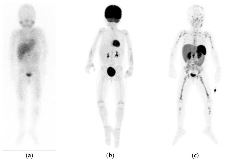
Figure 3.
Example of a patient with neuroblastoma, showing diffuse pathological but faint osteomedullary uptake on planar [123I]mIBG scintigraphy (a), without increased uptake on [18F]FDG positron emission tomography (PET) maximum intensity projections (MIP) (b), but extensive pathological osteomedullary uptake on [68Ga]Ga-DOTATATE PET MIP (c). Note the different physiological distribution between tracers.

