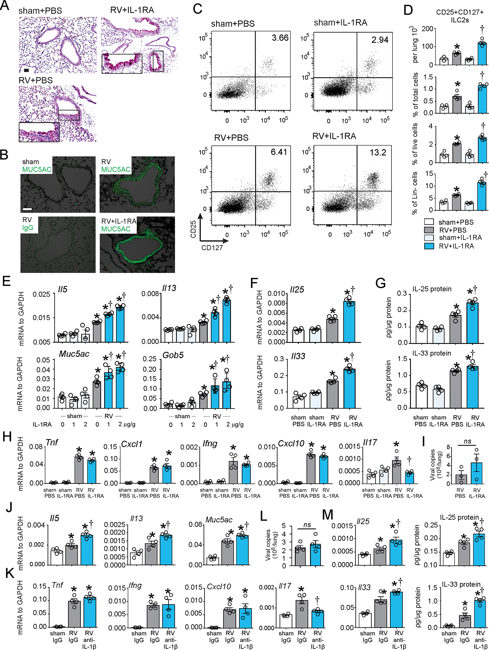
FIG 2. IL-1RA increased RV-induced ILC2 expansion and mucus metaplasia in 6-day old wild type mice.

Six day-old C57BL/6 mice were inoculated with sham or RV intranasally. IL-1RA or vehicle was given intraperitoneally one hour before RV infection, followed by a half dose of IL-1RA on day 1. The concentration of IL-1RA was 2 μg/g unless otherwise noted. PAS staining (A; bar=50 μm), Muc5ac immunofluorescence (B; bar=50 μm), lung lineage-negative CD25+ CD127+ ILC2s (C and D) and whole-lung mRNA and protein expression (E-H) were examined. Il33, Il17, Ifng, Tnf, Cxcl1 and Cxcl10 mRNA and IL-11 protein were examined at one day post-infection. Il25, Il5, Il13, Muc5ac and Gob5 mRNA IL-25 protein, PAS staining, Muc5ac immunofluorescence and lung ILC2s were examined 7 days post-infection. I. RV positive-strand RNA was assessed 24 h after infection, and presented as viral copy number in total lung. (N =3–4, mean±SEM, *different from sham, †different from RV, p<0.05, p<0.05, one-way ANOVA). J-M, 6-day old wild type C57BL/6 mice were inoculated with sham, RV, in combination with either isotype IgG control or anti-IL-1β Ab. Whole-lung mRNA (J, K) and vRNA (L) were measured using quantitative PCR, and whole lung IL-25 and IL-33 protein (M) were examined by ELISA. (N =4, mean±SEM, *different from WT sham, †different from WT RV, p<0.05, one-way ANOVA).
