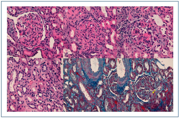
Figure 1.
(A) (10×) (B) (20×) (C) (40×) H.E: Glomeruli with endocapillary proliferation, observing collapsed vascular lumens and increased cellularity with isolated neutrophils (white arrow) and nuclear fragments (*). Thrombi are not observed; (D) (20×), HE: Tubules with isometric cytoplasmic vacuolization, (E) (10×) (F) (40×) Trichrome stain: erythrocytes in peritubular capillaries and solitary glomerular hyaline thrombus (yellow arrow).

