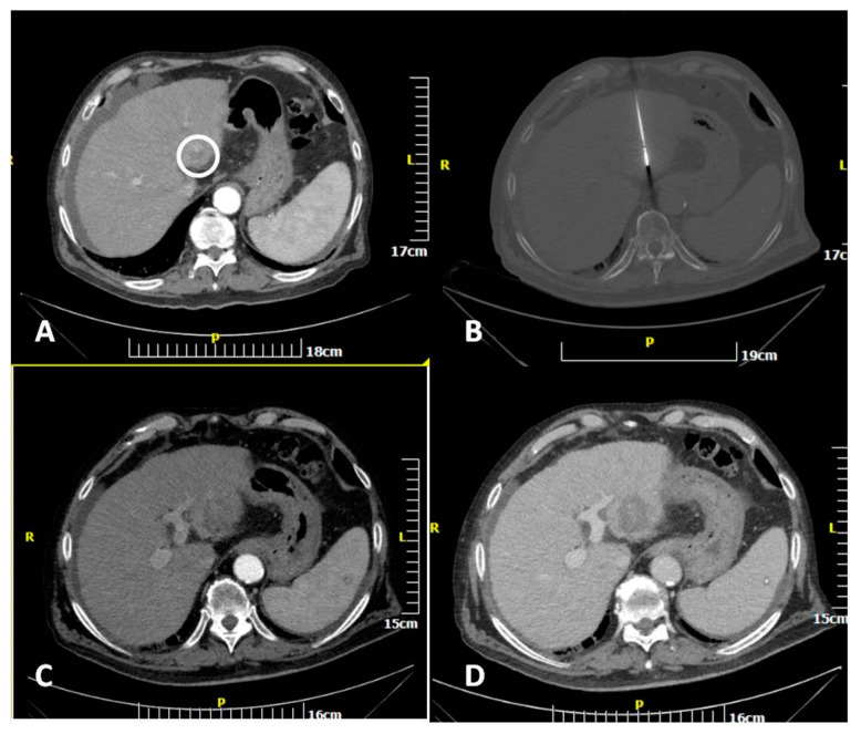
Figure 2.
CT scan of an 82-year-old man showing a hepatocellular cancer lesion. Enhanced transverse (A) CT image illustrates the lesion (white circle). Transverse CT image (B) illustrating the microwave antenna at the lesion level. Transverse CT images in arterial (C) and portal venous (D) phases illustrating the ablation zone.

