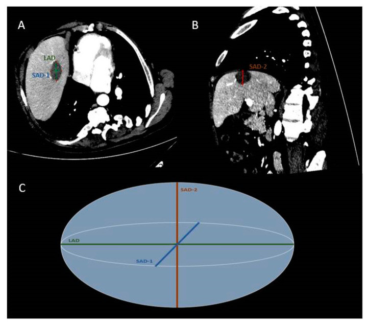
Figure 4.
CT scan of a 54-year-old man showing a colorectal cancer metastasis in liver segment VIII post microwave ablation (MWA). Enhanced transverse (A) and parasagittal (B) CT images. Simplified scheme of MWA zone (C). The short-axis diameters (SAD-1,-2) are orientated perpendicular to the long-axis diameter (LAD).

