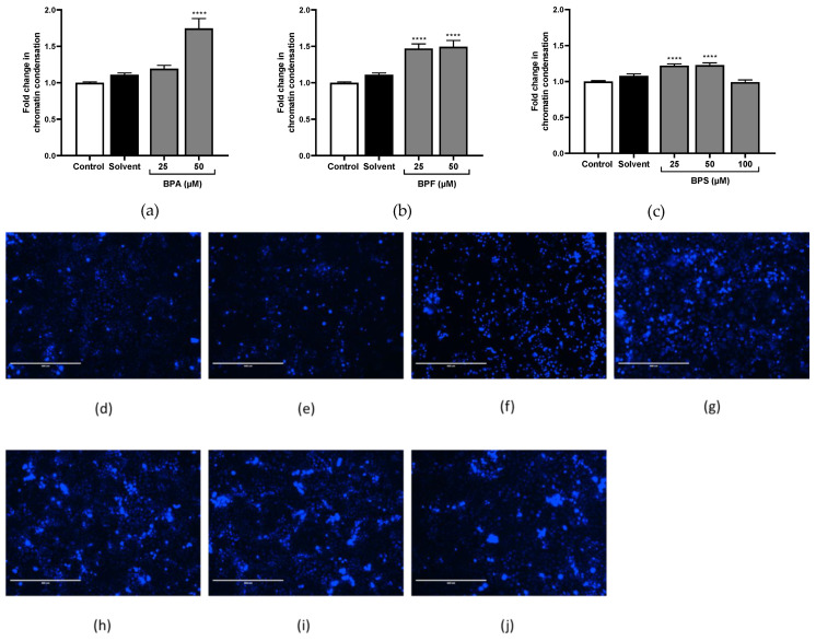
Figure 8.
Quantification and qualification of chromatin condensation using Hoechst 33,342 assay. (a–c) Chromatin condensation quantification was assessed after (a) BPA, (b) BPF, and (c) BPS incubation for 72 h in JEG-Tox cells. Data correspond to the mean ± SEM of four independent experiments. The significance threshold was **** p < 0.0001 compared to the control. (d–j) Chromatin condensation observation under fluorescence microscopy in JEG-Tox cells after incubation for 72 h with (d) the control, (e) solvent, (f) 50 μM of BPA, (g,h) 25 μM and 50 μM of BPF, respectively, and (i,j) 25 μM and 50 μM of BPS, respectively. The pictures are representative of three independent experiments and were captured under the same acquisition parameters.

