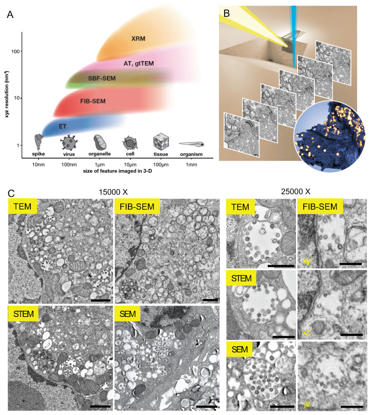
Figure 1.
Focused ion beam scanning electron microscopy (FIB-SEM) versus other electron microscopy (EM) approaches in imaging viruses and cells. (A) FIB-SEM occupies a crucial gap in a plot of typical resolutions achieved against the size of volumes imaged by volume EM (vEM) techniques. XRM: X-ray microscopy, AT: array tomography, gtTEM: grid-tape TEM, SBF-SEM: serial block face SEM, FIB-SEM: focused ion beam SEM, ET: electron tomography. (B) Schematic of FIB-SEM acquisition. Iterative gallium ion milling (blue beam) and scanning electron imaging (yellow beam) produce serial micrographs, which are aligned and segmented to reveal features such as infected cell membranes (dark blue) and viral particles (yellow spheres). (C) Representative images of SARS-CoV-2 infected Vero E6 cells taken by different EM imaging modalities. Images were acquired at ~15,000× (left) or ~25,000× (right) magnifications. TEM: transmission electron microscopy, STEM: scanning transmission electron microscopy, FIB-SEM xy, xz, yz: representative slices from orthogonal planes of the reconstructed volume. The raw FIB-SEM and SEM images were contrast inverted to match TEM and STEM micrographs visually. Scale bars: 1 µm (left), 500 nm (right).

