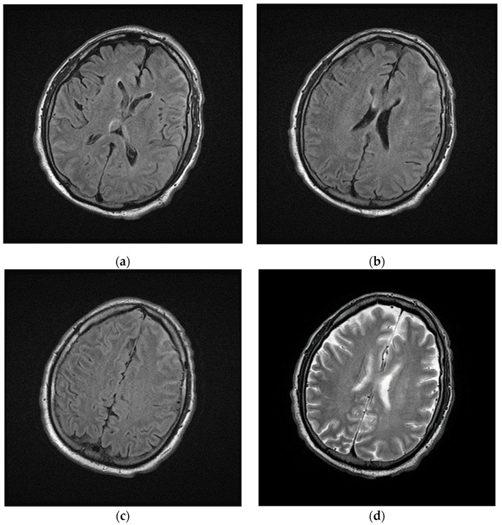
Figure 2.
Magnetic Resonance Imaging (MRI) of brain performed during intensive care. (a–c) Axial fluid-attenuated inversion recovery (FLAIR) images and (d) axial T2w image show cerebrospinal fluid hyperintensities in the left frontal lobe; in the same images no focal lesions of the cerebral parenchyma were detected.

