Figure 1.
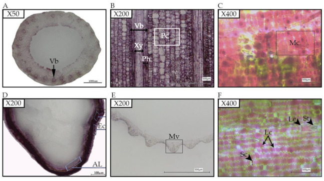
Anthocyanin pigmentation in different tissues of black rice; (A), a cross-section of stem showing vascular bundle (Vb) containing the anthocyanin; (B), leaf sheath longitudinal section showing a high amount of anthocyanin in parenchyma cells (Pc), xylem (Xy) and phloem (Ph); (C), the longitudinal section of ligule showing mesophyll cell (Mc) filled with the anthocyanin; (D), a cross-section of the caryopsis showing the aleurone layer (AL), the epidermal cells (Ec), and the transverse cells (Tc) colored by the anthocyanin; (E), a cross-section of a leaf blade with the midvein (Mv) containing the anthocyanin; (F), the longitudinal section of a leaf showing long cells (Lc) and silica cells (Sc) filled with anthocyanin, and stomate (st).

