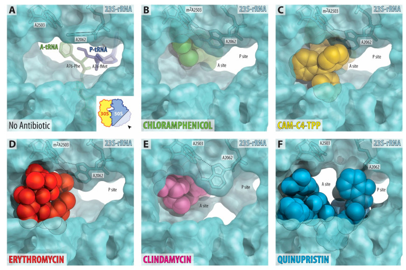
Figure 5.
Comparison of the binding sites of several antibiotics in the nascent peptide exit tunnel. (A) The lumen of the NPET in the drug-free 70S ribosome (PDB entry 4Y4P [32]). The view is from the wide-open part of the tunnel onto the PTC, as indicated by the inset. Aminoacylated nucleotides A76 of the A- and P-site tRNA are shown in green and dark blue, respectively. Note that nucleotide A2062 of the 23S rRNA is pointed toward the viewer and is not involved in Hoogsteen-edge base pairing with the nucleotide m2A2503 in the NPET wall. (B–F) Occlusion of the nascent peptide exit tunnel by chloramphenicol (B, green, PDB entry 6ND5 [2]), CAM-C4-TPP (C, yellow), macrolide erythromycin (D, red, PDB entry 6XHX [28]), lincosamide clindamycin (E, magenta, PDB entry 4V7V [4]), and type B streptogramin quinupristin (F, blue, PDB entry 4U26 [38]). Note that, unlike A-site binding chloramphenicol, the NPET-binding antibiotics, such as macrolides, lincosamides, or streptogramins B, as well as CAM-C4-TPP partially obstruct the lumen of the NPET. Also, note that, in the presence of ribosome-bound CAM-C4-TPP, there is enough space for a nascent peptide to pass through.

