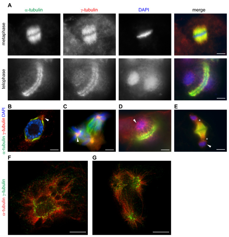
Figure 2.
γ-Tubulin localizes with mitotic microtubular arrays and a nuclear envelope in a cell cycle-dependent manner in acentrosomal plant cells. (A–G): Immunofluorescence labelling of γ-tubulin and α-tubulin in Arabidopsis cells—(A) γ-Tubulin localizes with microtubules of mitotic spindle and phragmoplast. (B–E): γ-Tubulin localization in cells treated with roscovitine—(B) γ-Tubulin forms condensated protrusions at polar regions in the vicinity of nuclei in cells arrested at G2/M (arrowhead); (C) γ-Tubulin foci in centers of chromosomal asters (arrowhead) of a multipolar spindle of Vicia faba; (D,E) γ-Tubulin is localized with minus ends of microtubules of aberrant phragmoplasts, often extending into the cytoplasm (asterisks) and patches of γ-tubulin are observed with newly formed nuclei (arrowhead); (F,G): STED (stimulated emission depletion) microscopy images of roscovitine- and taxol-treated cells of Arabidopsis. (time-gated continuous wave STED, 660 nm depletion laser, deconvolution by Huygens); (F) γ-Tubulin fibrillar arrays accumulate with nuclei in cells arrested at G2/M; (G) γ-Tubulin fibrillar arrays are enriched in the centers of chromosomal asters of multipolar mitosis. (A–E): Olympus Cell-R microscopy—(F,G): super-resolution Leica TCS STED 3X microscope. Scale bars: 5 µm (A–G).

