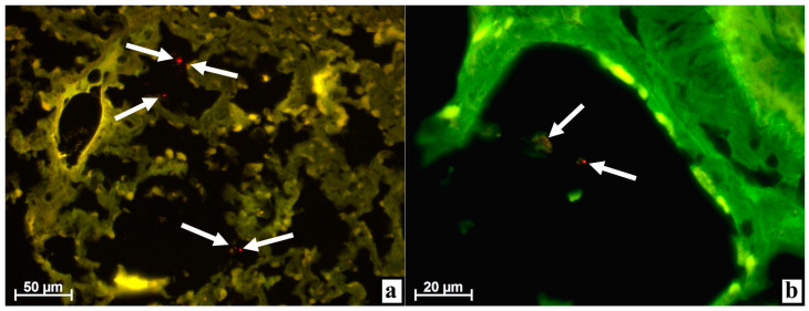
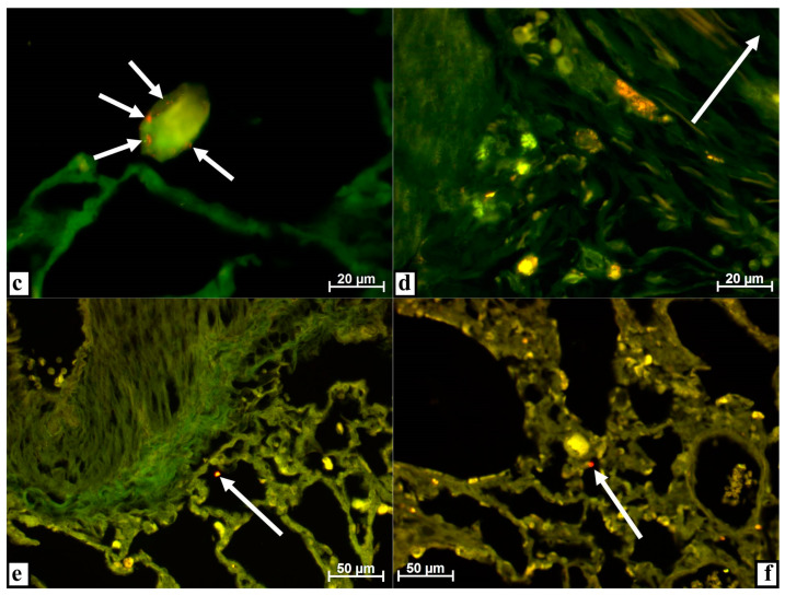
Figure 1.
Lungs of rabbits at various times after injecting extracellular vesicles derived from multipotent stromal cells (MSC EVs) into the tibial defect. Alignment of images obtained using Alexa 488 and rhodamine filters in the luminescent mode of the microscope. (a) After 3 days, numerous objects of very small size, dust-like, with bright fluorescence when using the rhodamine filter (arrows) were found in the alveoli. (b) In the alveolar lumen, objects with luminescence when applying the rhodamine filter were associated with a structureless substance (arrows) on day 3. (c) After 3 days, numerous objects with very bright fluorescence when using the rhodamine filter were enclosed in a homogeneous substance in the alveolus of the lung (arrows). (d) Cytoplasmic inclusions in large cells of various shapes have a red glow when applying the rhodamine filter in the lung parenchyma near a large vessel (the lumen is indicated by an arrow) on day 3. (e) By the 7th day, an object of about 5 μm in size (arrow) with intense fluorescence when using the rhodamine filter was found in the alveolus. (f) After 10 days, the alveolus contained an object with a diameter of 5–7 μm (arrow) with a very bright glow when a rhodamine filter was installed.


