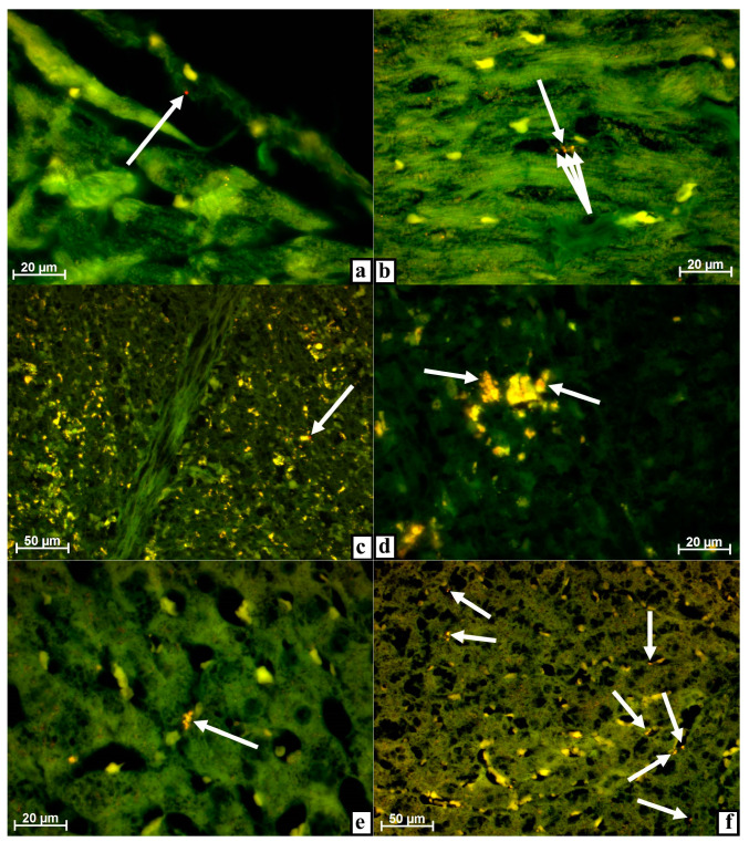
Figure 2.
Myocardium (a,b), spleen (c,d), and liver (e,f) of rabbits at different dates after damage to the tibia with the introduction of MSC EVs. Alignment of images obtained using Alexa 488 and rhodamine filters in the luminescent mode of the microscope. (a) On day 3, a very fine, dust-like object with strong fluorescence was located on the capillary endothelium (arrow) when a rhodamine filter was applied. (b) After 7 days, the capillary contained several very small, dust-like objects with a bright glow (arrow) when a rhodamine filter was installed. (c) After 3 days, a very small, dust-like object with a strong glow was noticeable close to a macrophage with intense autofluorescence located in the red pulp (arrow) when a rhodamine filter was used. (d) Individual inclusions in the cytoplasm of red pulp macrophages fluoresce with a noticeable red tint (arrows) by day 7 when a rhodamine filter was installed. (e) After 3 days, a macrophage in a sinusoid had a red tint in the glow of cytoplasmic inclusions (arrows) when a rhodamine filter was applied. (f) After 7 days, numerous macrophages were located along the sinusoids fluoresce with a clear red tint (arrows) when a rhodamine filter was installed.

