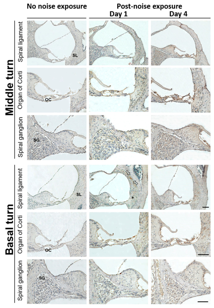
Figure 3.
Representative expression and distribution of HMGB1 in mouse cochlear tissues following noise exposure. HMGB1 immunohistochemical staining (brown color) was substantially increased on post-exposure day 1 in the spiral ligament of the basal turn. Arrows indicate HMGB1-positive staining cells, mainly localized in the type II cell region. On day 4, the HMGB1 was markedly expressed in the organ of Corti, spiral limbus, spiral ganglion, and spiral ligament of the cochlear basal and middle turns (n = 4 [refers to 4 cochleae from 4 different animals]). Scale bars = 50 μm. SL = spiral ligament; OC = organ of Corti; SG = spiral ganglion.

