Figure 6.
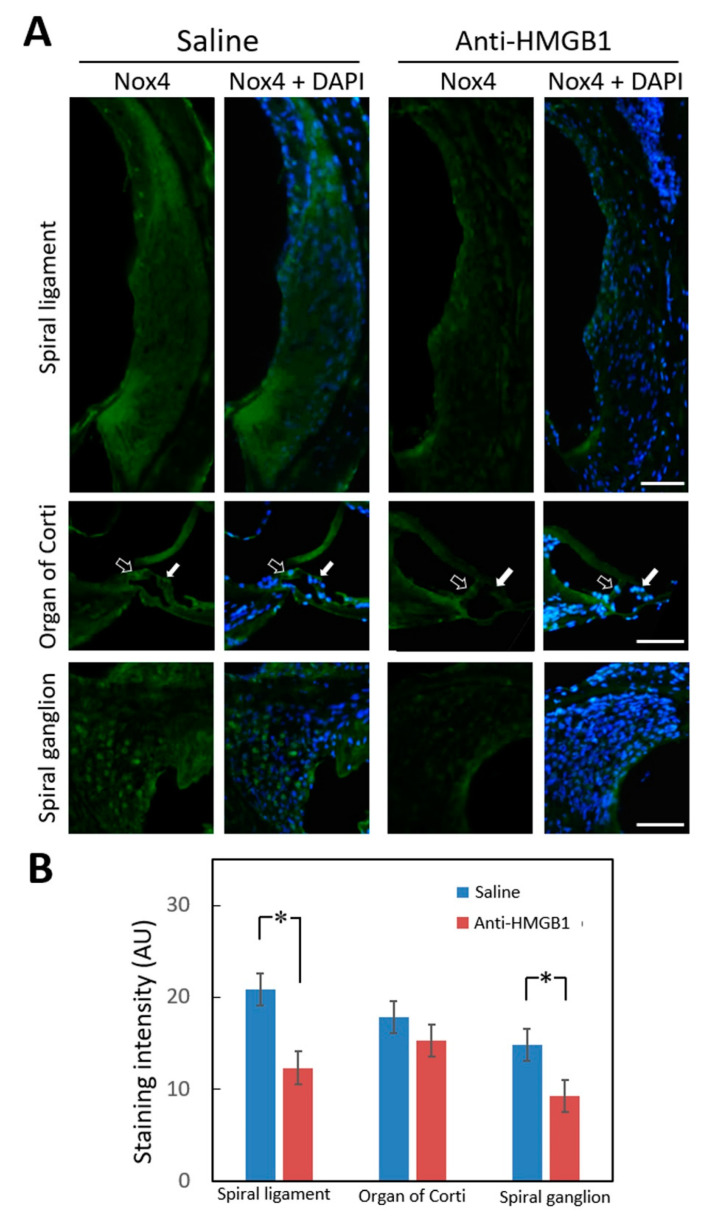
Immunohistochemical staining for NOX4 in the cochlea 4 days after noise exposure. (A) Representative staining of cochlear sections from mice pretreated with anti-HMGB1 antibodies or saline. Hollow arrows indicate inner hair cells and white arrows indicate outer hair cells. Labeling: Nox4 (green); DAPI (blue). (B) Histogram representations of the mean fluorescence intensity of Nox4 staining. Data are shown as the means ± SEM (n = 4 [refers to 4 cochleae from 4 different animals]) for each bar). Scale bars = 50 μm. * p < 0.05; Nox4 = NADPH oxidase 4; DAPI = 4,6-diamidino-2-phenylindole; SEM = standard error of the mean.

