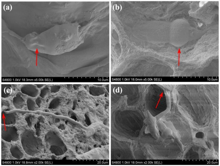
Figure 4.
Morphology of conidia and mycelial on branch under cryo-scanning electron microscope after 6 h of pathogen inoculation. (a,b) conidia on branches of resistant and susceptible cultivar, respectively; (c,d) mycelial on branches of resistant and susceptible cultivars, respectively. The red arrow in (a,b) represents the shrunk and expanded cell, respectively, while the red arrow in (c,d) represents the shrunk and natural mycelium, respectively.

