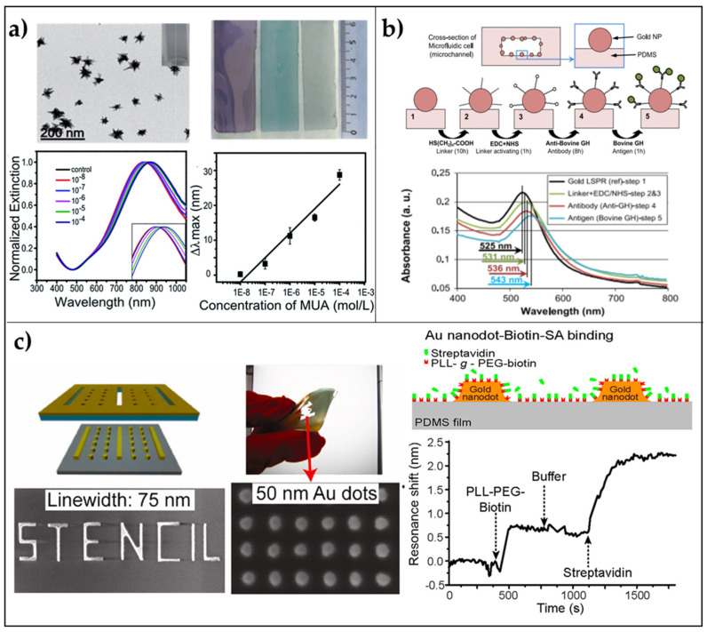
Figure 2.
2D arrays of AuNPs on PDMS flexible substrates. (a) Immobilization of AuNSs on PDMS: TEM images of gold nanostars (scale bar 200 nm); macroscopic morphology of PDMS/AuNSs platforms; extinction spectra of a PDMS/AuNSs film immersed in different concentrations of MUA in ethanol; LSPR shift versus MUA concentration. Reproduced from [71] with permission from Royal Society of Chemistry Pub. (b) In situ growth of AuNPs in a microfluidic chamber: Biosensing experiments performed by using the annealed microfluidic biosensor (400-2 cells) prepared from 2% aqueous solution of the gold precursor (48 h); cross-section of a microchannel and AuNPs in the channel; four steps of the biosensing protocol; a legend of the schematics; Au-LSPR corresponding to the four sensing steps, and LSPR band shift corresponding to different Ag concentrations. Reproduced from [72] Copyright (2013), with permission from Elsevier. (c) Patterning via Nanostencil Lithography: Schematics of biosensing experiments with nanodots on PDMS. Au nanodots and PDMS are biotin-functionalized, and then streptavidin binds to biotin. LSPR shift of an array of W = 75 nm and S = 50 nm Au nanodots on PDMS upon the addition of biotinylated molecules and streptavidin. The arrows indicate the addition of biomolecules and buffer rinsing. Adapted from [73]. Copyright (2012) with permission from American Chemical Society.

