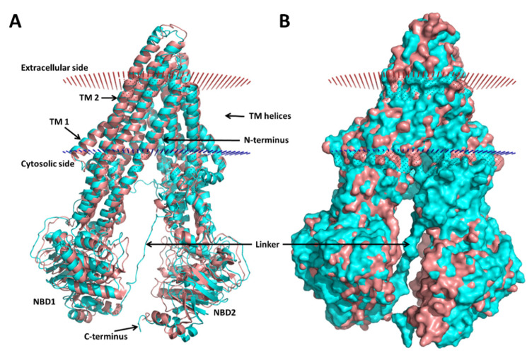
Figure 1.
Homology model 1 of the human P-glycoprotein (P-gp) superimposed with a crystal structure of the mouse P-gp (PDB ID: 4M1M). (A) Cartoon representation and (B) surface representation. The mouse P-gp is colored in salmon, and the model is colored in cyan; the cell membrane boundaries predicted by PPM 2.0 for the model are shown: the red dots represent the extracellular side of the membrane and the blue dots the cytoplasmic side. Several regions of the P-gp are indicated: the nucleotide-binding domains (NBDs) where the ATP binds, the transmembrane (TM) α-helices 1 and 2 (TM 1 and 2); the linker between the two halves (ca. residues 631–684 in the human P-gp numeration), which is missing in the crystallographic structures.

