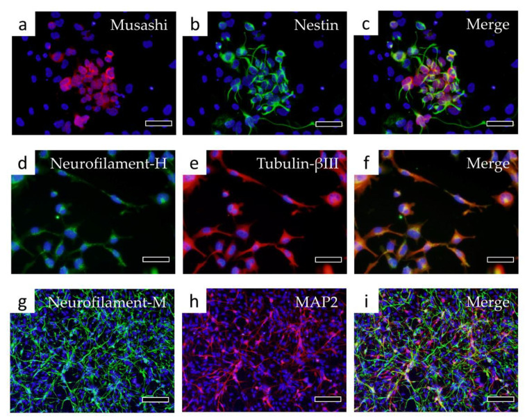
Figure 6.
Immunostaining of cells differentiated into nerve cells. Fluorescent immunostaining was performed with the following markers of neural stem/progenitor cells: (a) Musashi, (b) Nestin, (c) Merge of Musashi and Nestin. Immunostaining was then performed with the following markers of nerve cells: (d) Neurofilament-H, (e) Tubulin-βIII, (f) Merge of Neurofilament-H and Tubulin-βIII, (g) Neurofilament-M, (h) MAP2, (i) Merge of Neurofilament-M and MAP2. Bars in panels (a–f): 50 μm, bars in panels (g–i): 100 μm.

