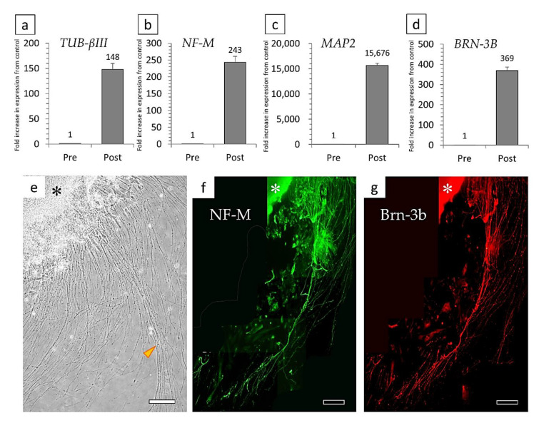
Figure 8.
Relative semi-quantitative analysis of the gene expressions of (a) TUBULIN-βIII, (b) NEUROFILAMENT-M, (c) MAP2, and (d) BRN-3B in pre/post-differentiated cells. (e) An adhesive culture of cell aggregates (*) formed in suspension culture resulted in very long neurite outgrowth (arrowhead). (f,g) Fluorescent immunostaining results for markers of retinal ganglion cells: (f) Neurofilament-M, and (g) Brn-3b. Bars: 200 μm.

